Machine Learning Methods For Knee Feature Extraction From Mr Images
From this perspective feature extraction or. Gibbs-ringing artifact in MR images is caused by insufficient sampling of the high frequency data.

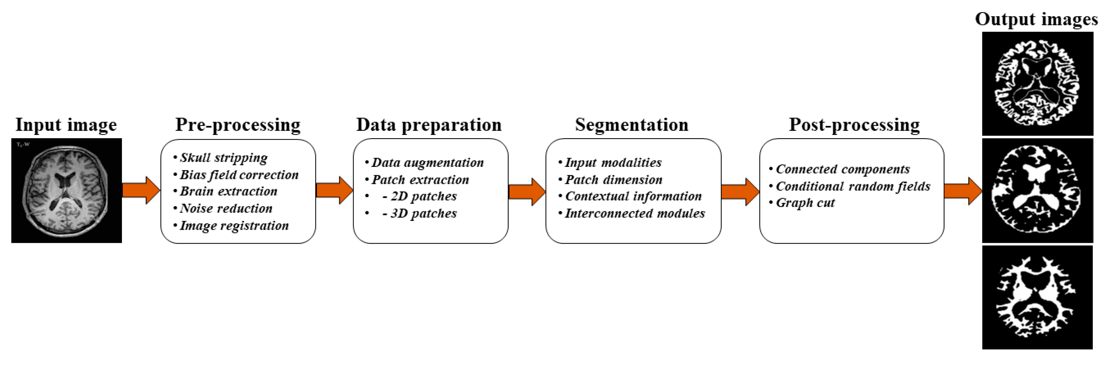
Preparing Medical Imaging Data For Machine Learning Radiology
The experimental results have presented with proposed approach.
Machine learning methods for knee feature extraction from mr images. In this paper feature extraction method is proposed and performed on medical images which CT scan Cancer datasetss. The processed feature set and original raw feature set were severed as input to four. Deep learning that is a subfield of machine learning concerned with algorithms inspired by the structure and function of the brain sets an alternative architecture by shifting the burden of feature engineering the process of transforming raw data into features to the underlying learning system.
Rheumatoid Arthritis RA is considered as a chronic disease that occurs due to progressive changes in the structural properties of knee tissues. To develop a machine learning approach using convolutional neural network for reducing MRI Gibbs-ringing artifact. Deep learning will soon help radiologists make faster and more accurate diagnoses.
Then images with same severity will have. Feature extraction starts with feature detection where features in an image are local regions of pixels that include an interesting part of a microstructure such as a corner edge or blob-like object. These data support the feasibility of multiinstitutional classification of radiologic imaging text reports with a single machine learning classifier without requiring institution-specific training data.
The main advantage of this method is the ability to learn the adaptive image. We have computed the Cartilage Damage Index CDI information from 36 informative locations on tibiofemoral compartment from 3D MR imaging reconstruction and used PCA analysis to process the feature set. This paper explored the hidden biomedical information from knee magnetic resonance MR images for osteoarthritis OA prediction.
In feature extraction combination of Modified Multi-Texton Histogram MMTH and Multi-Texton Microstructure Descriptor MTMD is used and then Gray Level Co-occurrence Matrix GLCM and Gray Level Run Length Matrix GLRLMare used to extract the feature from the image to. Stanford ML Group led by Andrew Ng works on important problems in areas such as healthcare and climate change using AI. To do this we cut the X-ray image vertically divide the single image into two pictures of the same size and do cluster analysis on both of them.
A Novel Method to Predict Knee Osteoarthritis Progression on MRI Using Machine Learning Methods. Furthermore the machine learning classifier performed well on free-text knee MRI reports from another institution. Large number of algorithms has been proposed for the extraction of features from knee MRI images.
Image sourceOver the last decade the ability of computer programs to extract information from images. Paper gives the impact of feature extraction that used in a deep learning technique such as Convolutional Neural Network CNN. Statistical structural spectral filtering histograms transformation and many more methods are used for texture feature extraction.
The global features capture the. Feature extraction is the first step in the process of classifying micrographs. Knee the most intuitive detection of OA is to calculate the distance of the bones between edge.
Existing methods exploit smooth constraints to reduce intensity oscillations near sharp edges at the cost of blurring details. However this study is specifically aimed to apply machine-learning algorithms with feature extraction approach. In particular cartilage thickness bone density meniscus volume and synovial fluid composition are the key causes for.
For those wishing to enter the field. In this paper we present an iterative multi-class learning method to. There are vast applications of deep convolutional neural network DCNN and machine-learning algorithms in medical imaging problems 32 3435363738.
The automatic segmentation of human knee cartilage from 3D MR images is a useful yet challenging task due to the thin sheet struc-ture of the cartilage with di use boundaries and inhomogeneous intensi-ties. This study explored the hidden biomedical information from knee MR images for osteoarthritis prediction. Machine Learning Methods for Knee Feature Extraction from MR Images Abstract.
Subsequently the MRNet challenge was also announced. The processed feature set and original raw feature set were severed as input to four machine learning methods artificial neural network ANN support vector. Texture analysis serve as a base for various feature description.
Last year they released a knee MRI dataset consisting of 1370 knee MRI exams performed at Stanford University Medical Center. Du Y Almajalid R Shan J Zhang M. NLEpy -- baseline method of this thesis which do not depend on any machine learning model PCApy -- Principle Component AnalysisPCA of all the extracted features Plotpy -- Plot the distribution of whole the features which is compressed into three dimension after PCA.

Dose Evaluation Of Mri Based Synthetic Ct Generated Using A Machine Learning Method For Prostate Cancer Radiotherapy Medical Dosimetry
Potential Use Of Deep Learning Techniques For Postmortem Imaging Springerlink

Artificial Intelligence And Machine Learning In Spine Research Galbusera 2019 Jor Spine Wiley Online Library

Machine And Deep Learning Methods For Radiomics Avanzo 2020 Medical Physics Wiley Online Library

A Machine Learning Approach For The Identification Of New Biomarkers For Knee Osteoarthritis Development In Overweight And Obese Women Osteoarthritis And Cartilage

Machine And Deep Learning Methods For Radiomics Avanzo 2020 Medical Physics Wiley Online Library
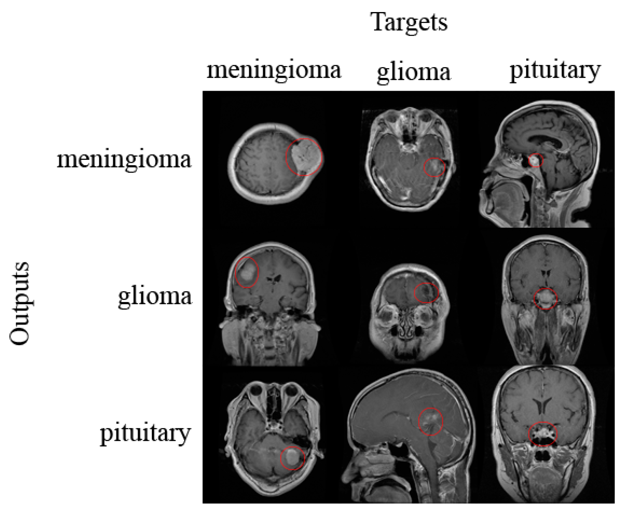
Applied Sciences Free Full Text Classification Of Brain Tumors From Mri Images Using A Convolutional Neural Network Html
It S A No Brainer Deep Learning For Brain Mr Images By Henrik Marklund Stanford Ai For Healthcare Medium
Imaging Applications Of Artificial Intelligence Healthmanagement Org
Cancers Free Full Text Optimizing Neuro Oncology Imaging A Review Of Deep Learning Approaches For Glioma Imaging Html

Accuracy Of Deep Learning To Differentiate The Histopathological Grading Of Meningiomas On Mr Images A Preliminary Study Banzato 2019 Journal Of Magnetic Resonance Imaging Wiley Online Library

Machine Learning And Deep Learning In Medical Imaging Intelligent Imaging Journal Of Medical Imaging And Radiation Sciences
Imaging Applications Of Artificial Intelligence Healthmanagement Org

Deep Learning In Radiology An Overview Of The Concepts And A Survey Of The State Of The Art With Focus On Mri Mazurowski 2019 Journal Of Magnetic Resonance Imaging Wiley Online Library
It S A No Brainer Deep Learning For Brain Mr Images By Henrik Marklund Stanford Ai For Healthcare Medium

Machine Learning For Detecting Early Infarction In Acute Stroke With Non Contrast Enhanced Ct Radiology

Deep Learning In Radiology An Overview Of The Concepts And A Survey Of The State Of The Art With Focus On Mri Mazurowski 2019 Journal Of Magnetic Resonance Imaging Wiley Online Library
Sensors Free Full Text Mri Segmentation And Classification Of Human Brain Using Deep Learning For Diagnosis Of Alzheimer S Disease A Survey Html
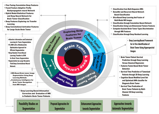
Brain Sciences Free Full Text Brain Tumor Analysis Empowered With Deep Learning A Review Taxonomy And Future Challenges Html
Post a Comment for "Machine Learning Methods For Knee Feature Extraction From Mr Images"